
HSP70
;Purple line: Antigen (10ng); Blue line: Antigen (50 ng); Red line:Antigen (100 ng)")
图片
- Elisa
Black line: Control Antigen (100 ng);Purple line: Antigen (10ng); Blue line: Antigen (50 ng); Red line:Antigen (100 ng)
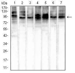 Western Blot
Western blot analysis using HSP70 mouse mAb against NIH/3T3 (1), Hela (2), HepG2 (3), Hek293 (4), COS-7 (5), A549 (6), and Jurkat (7) cell lysate.
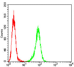 Flow cytometric
Flow cytometric analysis of Hela cells using HSP70 mouse mAb (green) and negative control (red).
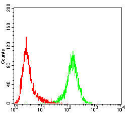 Flow cytometric
Flow cytometric analysis of Jurkat cells using HSP70 mouse mAb (green) and negative control (red).
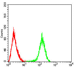 Flow cytometric
Flow cytometric analysis of Raji cells using HSP70 mouse mAb (green) and negative control (red).
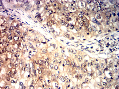 Immunohistochemistry
Immunohistochemical analysis of paraffin-embedded human breast cancer tissues using HSP70 mouse mAb with DAB staining.
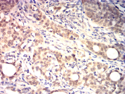 Immunohistochemistry
Immunohistochemical analysis of paraffin-embedded human cervical cancer tissues using HSP70 mouse mAb with DAB staining.
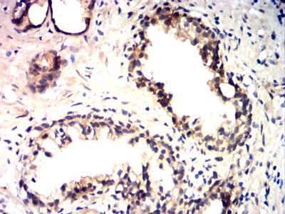 Immunohistochemistry
Immunohistochemical analysis of paraffin-embedded human prostate cancer tissues using HSP70 mouse mAb with DAB staining.
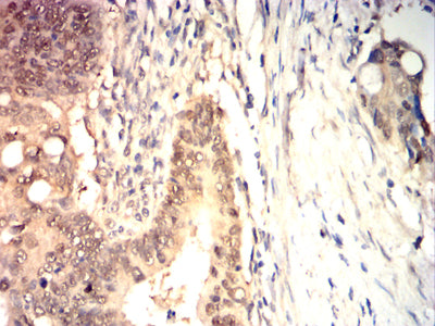 Immunohistochemistry
Immunohistochemical analysis of paraffin-embedded human rectal cancer tissues using HSP70 mouse mAb with DAB staining.
