
ATP1A1
;Purple line: Antigen (10ng); Blue line: Antigen (50 ng); Red line:Antigen (100 ng)")
图片
- Elisa
Black line: Control Antigen (100 ng);Purple line: Antigen (10ng); Blue line: Antigen (50 ng); Red line:Antigen (100 ng)
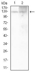 Western Blot
Western blot analysis using ATP1A1 mouse mAb against Hela (1) and A431 (2) cell lysate.
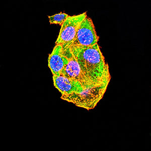 Immunofluorescence
Immunofluorescence analysis of Hela cells using ATP1A1 mouse mAb (green). Blue: DRAQ5 fluorescent DNA dye. Red: Actin filaments have been labeled with Alexa Fluor- 555 phalloidin. Secondary antibody from Fisher (Cat#: 35503)
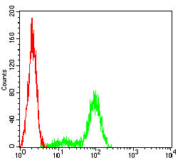 Flow cytometric
Flow cytometric analysis of Hela cells using ATP1A1 mouse mAb (green) and negative control (red).
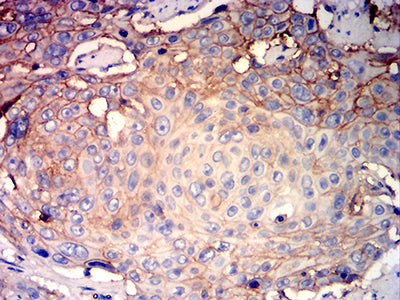 Immunohistochemistry
Immunohistochemical analysis of paraffin-embedded human esophageal cancer tissues using ATP1A1 mouse mAb with DAB staining.
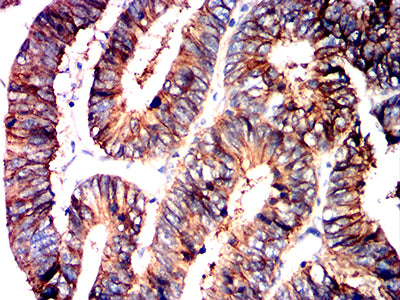 Immunohistochemistry
Immunohistochemical analysis of paraffin-embedded human rectum cancer tissues using ATP1A1 mouse mAb with DAB staining.
